There’s no substitute for methodical study when you examine horse locomotion, because analyzing the four natural gaits, hoof-sequence patterns, and biomechanics lets you detect lameness early, refine your movement-quality assessment, and elevate athletic performance through conformation-informed training and slow-motion analysis.
Key Takeaways:
- Horse locomotion is organized into four natural gaits—walk, trot, canter, gallop—with distinct hoof‑sequence patterns (walk: four‑beat lateral sequence; trot: two‑beat diagonal; canter: three‑beat with suspension; gallop: four‑beat with extended suspension) plus multiple ambling gaits (pace, rack, tölt) characterized by lateral coupling and altered duty factors that change center‑of‑mass trajectories and energy cost.
- Biomechanics-driven assessment focuses on temporal (stride time, stance/flight phases, limb phasing) and spatial (stride length, limb excursion, joint angles) metrics, plus kinetic indicators (peak vertical ground reaction force, impulse). Conformation and musculoskeletal geometry modulate leverage, joint loading, and propulsion, directly affecting movement quality and performance potential.
- Objective gait analysis (high‑speed video, slow‑motion kinematics, force plates, IMUs) enhances lameness detection and training/rehab decision‑making by quantifying asymmetry, compensatory patterns, and gait transitions; interpreting these metrics enables targeted interventions for athletic optimization and injury risk reduction.
Four Natural Gaits Explained
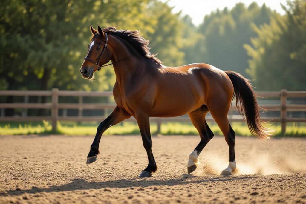
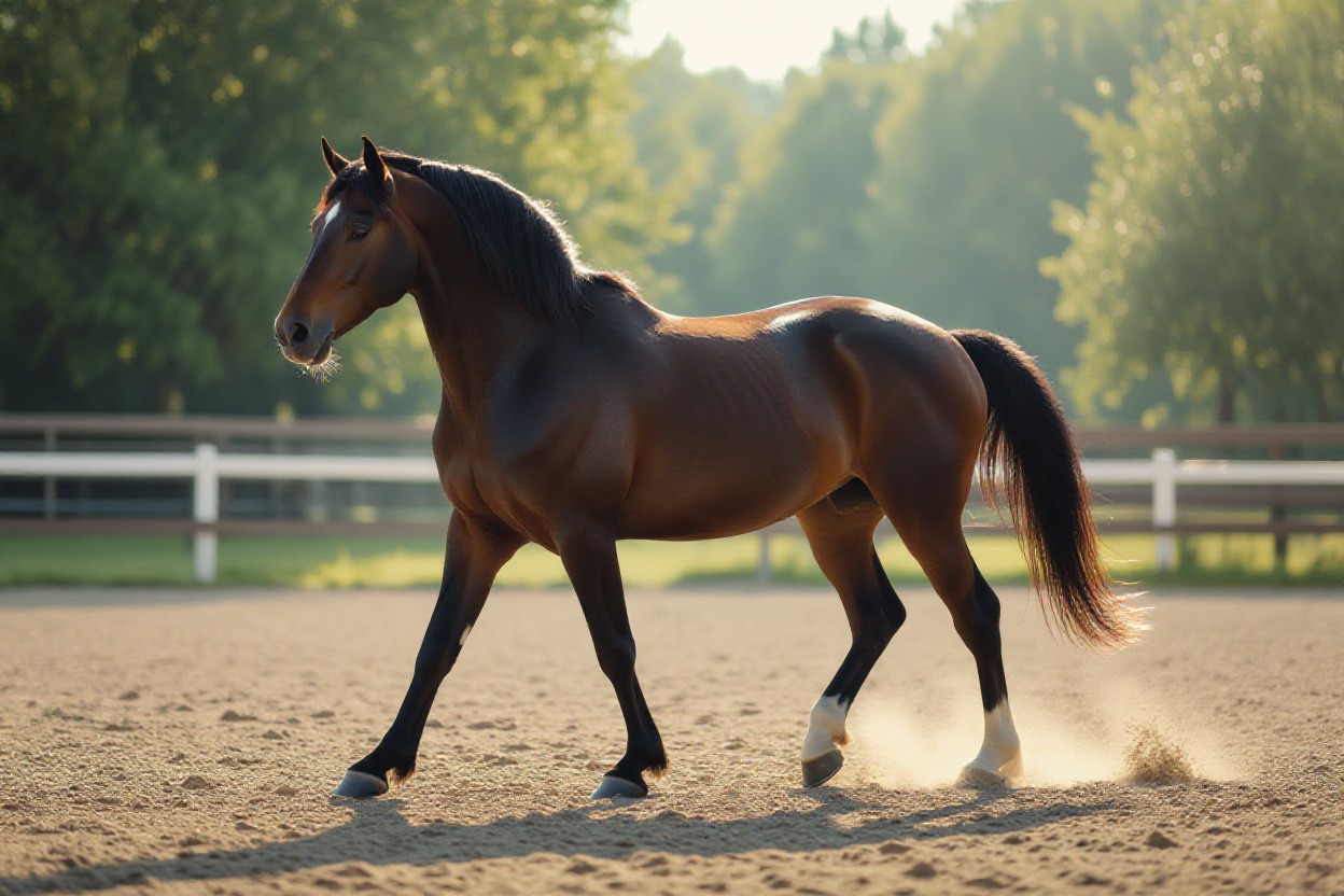
For detailed analysis of horse locomotion, you should compare the four natural gaits by biomechanics, hoof sequence and functional use: walk, trot, canter and gallop. Expect speed ranges from ~3–4 mph at the walk to race speeds of 40–45 mph at the gallop; you’ll use the trot most for lameness detection and the canter for balance and collection work. Each gait shows distinct ground-reaction force patterns and suspension phases that determine performance and injury risk.
Walk
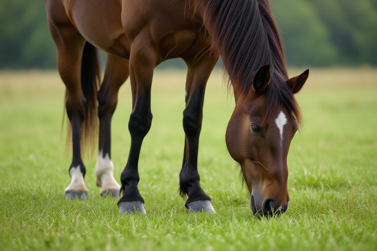
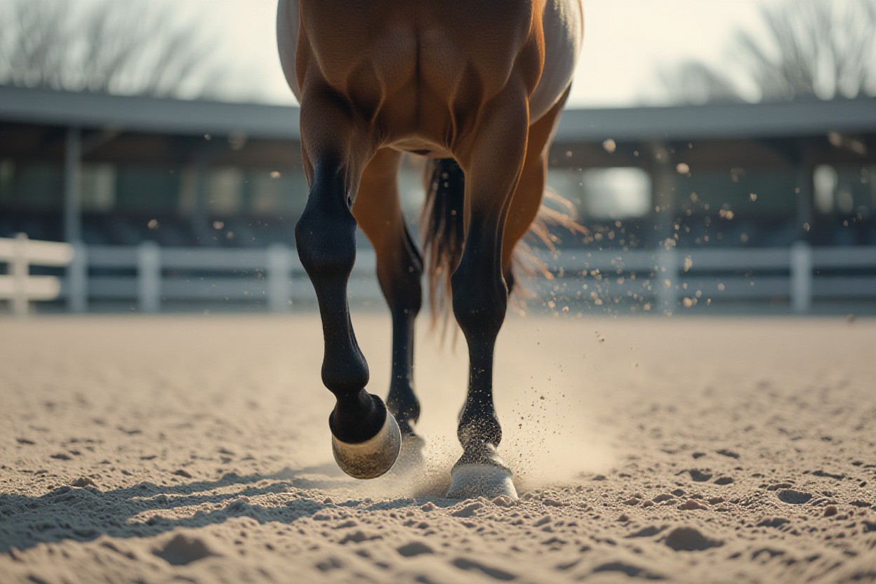
The walk is a true four-beat gait with an even hoof sequence and speeds around 3–4 mph; you’ll see each foot strike separated by roughly equal intervals, giving maximal stability and minimal concussion. Use the walk for rehabilitation, low-intensity conditioning and conformation assessment, but watch for stumbling on uneven or slippery ground since the walk’s slow cadence still exposes you to trip-related falls.
Trot
The trot is a two-beat diagonal gait used widely for training and diagnostics, typically at 8–13 mph with a stride rate near 90–120 strides per minute; you’ll see simultaneous diagonal limb pairs with a brief suspension phase, making it sensitive to asymmetry and ideal for detecting mild lameness.
In slow-motion analysis the trot magnifies left–right differences: head nods, hip hike and altered ground-reaction curves reveal fore or hind lameness. You should record at 120–240 fps to quantify stride-phase timing; subtle 5–10% force reductions between diagonals often indicate pathology, so the trot is your primary biomechanical screening gait.
Canter
The canter is a three-beat, asymmetrical gait (lead-dependent) typically between 10–17 mph; you’ll note the sequence—trailing hind, diagonal pair, leading fore—followed by suspension, which tests balance and collection. Use the canter to evaluate lead preference, impulsion and the effect of conformation on transverse rotation, but beware that cross-cantering signals poor balance and increases fall risk at speed.
Mechanically, the canter shifts peak vertical force to the leading forelimb and increases rotational moments around the spine; you should assess lead changes and suspension duration, as 20–30% differences in limb loading across leads can predict performance limitations or predisposition to soft-tissue injury.
Gallop
The gallop is an extended, four-beat gait with a long suspension phase and top speeds for race breeds of 40–45 mph; you’ll observe large stride lengths (often 6–8 m in Thoroughbreds) and the highest ground-reaction forces, making it the most athletically demanding and the gait with the greatest risk of catastrophic limb injury.
At the gallop, peak forces and repetitive loading drive fatigue and injury risk: you should monitor stride length, frequency and track surface effects, since a 5–10% reduction in stride length or abrupt asymmetry often precedes breakdowns; conditioning, conformation and footing jointly determine whether a horse sustains elite sprinting loads safely.
Ambling & Specialty Gaits
Among ambling and specialty patterns in horse locomotion you’ll encounter lateral and intermediate rhythms—rack, tölt, foxtrot and pace—that prioritize rider comfort and specific athletic tasks. You should note they differ in beat structure (two- to four-beat), typical speeds (trail amblers 6–12 mph; some specialty pacers/racers far faster) and how conformation channels forces through limbs, affecting gait symmetry, performance and lameness detection. Pay attention to cadence, stride length and consistency when assessing movement quality.
Foxtrot
The foxtrot is a four-beat, diagonal “broken trot” where the forefoot lands slightly before the diagonal hind, producing an exceptionally smooth, ground-covering ride favored by Missouri Fox Trotters; you’ll commonly see comfortable cruising speeds of about 6–12 mph. Observe long, elastic strides and a relaxed back to identify true foxtrot mechanics; subtle limb asymmetries are often masked, so evaluate both straight lines and circles for latent lameness.
Pace
Pace is a lateral two-beat gait with right and left fore–hind pairs striking nearly together, giving high straight-line efficiency but greater lateral instability; recreational pacers travel 10–25 mph while Standardbred racing pacers can reach elite mile times around 1:48 (~33–34 mph). When you assess pacing, check for limb interference, asymmetric loading and how turns increase concussion on the outside limbs—these factors drive both performance limits and injury risk.
Biomechanically, pacing produces synchronous lateral loading that raises torsional stresses across fetlocks and suspensory apparatus, so you should monitor for early signs like uneven hoof wear, widening stride variance or increased fetlock descent. Training interventions such as targeted conditioning, corrective shoeing and, in harness contexts, calibrated hobbles alter kinematics; note that pacing can both mask subtle soreness in straight lines and accelerate lateral limb injuries during high-speed turns.
Biomechanics of Horse Movement
In horse locomotion you must consider how mass, limb geometry and elastic tissues interact: peak vertical ground reaction forces reach ~2–3× body weight per limb depending on gait, and stride speeds range from ~1.5 m/s at the walk to >15 m/s in elite gallops. Tendons like the superficial digital flexor act as elastic springs, reducing metabolic cost, while limb phase timing and hoof placement determine stability and asymmetry; asymmetric loading often signals early lameness or performance loss.
Joint Function
You assess joint function by watching contributions from the shoulder and elbow (shock absorption and protraction) versus the stifle and hock (power generation). The fetlock’s large extension stores elastic energy but also concentrates load on the suspensory apparatus; excessive fetlock extension or repeated high peak loads increases risk of tendon and sesamoid injury. Gait-specific joint angles and timing alter force transmission—altered stifle extension at trot commonly precedes hindlimb lameness.
Muscle Engagement
You observe major muscle groups coordinating force and control: gluteals and hamstrings provide rear propulsion, longissimus dorsi stabilizes the trunk, and digital flexors control distal limb swing and hoof placement. Sprint-type horses often show higher proportions of fast-twitch fibers (up to ~60–70%) in proximal muscles, enhancing short-duration power; well-developed gluteal mass directly improves acceleration and stride length.
Electromyography and training studies show you how timing and fiber-type shifts affect performance: gluteus medius activity typically peaks in late stance to drive hock extension, while the longissimus fires across stance to resist pitching. Conditioning increases cross-sectional area and can shift fiber phenotype toward more fast-oxidative capacity in interval-trained horses. Monitor fatigue-induced timing shifts closely, since altered activation patterns are an early marker of decreased performance and higher injury risk.
Assessing Movement Quality
Assess movement quality by combining objective metrics—stride length, cadence, joint angles—with subjective reads of impulsion, balance and suspension. You should use high-speed video, inertial sensors and force-plate data to quantify deviations. Watch for head bobbing, shortened stride, uneven hoof flight and loss of suspension, as these often signal pain or mechanical restriction. Integrate conformation, gait-specific biomechanics and performance goals so your assessment of horse locomotion differentiates normal variation from pathology.
Symmetry and Rhythm
Evaluate footfall timing and phase durations: walk typically 90–120 steps/min, trot 120–140 strides/min, canter 70–90 strides/min and gallop 150–180 strides/min. Asymmetry in timing or vertical displacement exceeding ~5% of stride time or ~6 mm of head/hip motion is a red flag for lameness. Use slow-motion and sensor traces to quantify phase shifts; consistent unevenness across surfaces or speeds points to underlying injury or compensation.
Flexibility and Reach
Flexibility and reach determine stride length and athletic scope: typical stride lengths approximate trot 2.5–3.5 m, canter 3.5–5.5 m, gallop 5.5–7.0 m. Increased shoulder protraction and hindlimb retraction yield longer, more effective strides; conversely, restricted range from joint pain, tight musculature or poor shoeing produces reduced reach and loss of performance. You must inspect scapulohumeral, fetlock and pelvic ROM when diagnosing limited reach.
Assess reach on the move: perform in-hand straight-line and lunge tests at multiple speeds, then record at 240–500 fps or use IMU sensors to measure joint angles and protraction distance. When you detect a ~10° loss of shoulder flexion or diminished fetlock extension, expect roughly a 5–10% drop in measurable stride length. Targeted physiotherapy, farriery and conditioning often restore ROM; early intervention improves recovery and prevents chronic compensation.
Lameness Detection
Signs of Lameness
You should watch for asymmetric footfall patterns, a shortened stride, and compensatory changes—head nod on the trot often signals a forelimb issue, while hip hike or reduced hind propulsion indicates hindlimb pain. Palpable heat, swelling, focal pain on hoof testers, and altered hoof wear are common. Grade lameness on the AAEP 0–5 scale, with non-weight-bearing or sudden severe lameness demanding immediate action to prevent further damage.
Diagnostic Methods
Begin with a controlled trot-up and static palpation, then use flexion tests and hoof testers to provoke signs. Employ diagnostic analgesia (perineural and intra-articular blocks) in a distal-to-proximal sequence to localize pain. Follow with imaging—radiographs, ultrasound, CT or MRI for the foot—and gait quantification tools like high-speed video and force plates to objectify asymmetry.
You should sequence tests: start noninvasive, progress to blocks, then targeted imaging based on block results. Force plates (sample rates ~1,000 Hz) and IMUs pick up subclinical asymmetries invisible to the eye, while scintigraphy helps with multifocal or early bone lesions. For example, a performance horse with intermittent forelimb lameness may respond to a palmar digital block, directing you to MRI of the distal phalanx or navicular region to confirm pathology before treatment.
Common Gait Disorders
You will encounter a range of disorders that disrupt normal horse locomotion: laminitis (often affecting the forefeet and risking coffin bone rotation >10°), navicular syndrome producing short-strided forelimb lameness, osteoarthritis as the leading cause of chronic lameness in older athletes, tendon injuries (SDFT lesions common in race/athletic horses), and neurologic conditions such as cervical vertebral myelopathy. Use slow-motion and sensor data to detect subtle asymmetries; gait changes of >3–5% stride difference often indicate pathology requiring prompt evaluation.
Causes and Treatment
You should link causes to tailored treatments: metabolic issues (EMS, PPID) call dietary control and medical management, traumatic or overload injuries need rest and staged rehabilitation, and joint degeneration benefits from NSAIDs, intra-articular therapies, or arthroscopic debridement. For laminitis, immediate cryotherapy and offloading are priorities and, in severe cases, soft tissue surgery or deep digital flexor tenotomy may be considered. Always coordinate farriery, medications, and targeted therapies with your veterinarian and farrier.
Management Strategies
You must prioritize prevention and monitoring: schedule farriery every 4–6 weeks, implement controlled exercise programs, and apply early detection methods such as high-speed video or inertial sensors to quantify asymmetry. Emphasize regular body condition scoring and metabolic screening to lower laminitis risk. Early intervention and objective tracking dramatically reduce progression to irreversible damage and improve return-to-performance outcomes.
You can operationalize management by setting measurable targets: use IMUs to track stride symmetry weekly and flag >3% asymmetry, maintain turnout and graded water treadmill or hill work for 6–12 weeks during rehab, and employ shoeing solutions like egg-bar shoes for laminitic horses or heel wedges for palmar pain. Combine diagnostics (radiographs, ultrasound) with therapies such as PRP, shockwave, and a 8–12 week progressive loading plan to optimize tissue remodeling and performance restoration.
Summing up
Following this, you will be able to interpret horse locomotion across walk, trot, canter, gallop and ambling gaits, assess hoof‑sequence patterns, detect lameness, and evaluate how conformation and biomechanics influence your horse’s performance using slow‑motion analysis and movement‑quality metrics.
FAQ
Q: What are the primary gaits and their biomechanical and hoof-sequence characteristics in horse locomotion?
A: Horse locomotion is classically categorized into a set of natural gaits with distinct kinematic and kinetic signatures. The four standard gaits are the walk, trot, canter, and gallop; additionally, ambling gaits (pace, rack, tölt, running walk, stepping pace) occur in certain breeds or as trained variations. Each gait can be described by footfall sequence, beat (number of distinct contacts per stride), limb phasing, duty factor (percentage of stride time a foot spends on the ground), center-of-mass (CoM) motion, and presence or absence of suspension phases.
Walk: A four-beat, lateral-sequence gait with no full suspension phase at normal speed. Footfall pattern generalization: hind limb ipsilateral fore limb, then contralateral hind, then contralateral fore (e.g., RH → RF → LH → LF, depending on which limb starts). Duty factor > 0.5 for each limb, producing periods where three limbs bear weight simultaneously. Biomechanically the walk is an inverted pendulum-like transfer of CoM, with vaulting over a stiffening limb that minimizes vertical CoM oscillation and optimizes stability. Energy exchange between potential and kinetic energy is high; limb compliance, shoulder reach, and pelvic rotation set stride length.
Trot: A two-beat, diagonal gait (RH+LF, LH+RF) with a distinct suspension phase between diagonal contacts. Duty factor typically < 0.5 as speed increases, producing a measurable aerial phase. The trot uses spring-mass mechanics dominated by elastic energy storage and return in the superficial digital flexor tendon, suspensory apparatus, and muscle-tendon units. Diagonal pairing produces symmetrical vertical GRF (ground reaction force) patterns in sound animals and convenient diagnostic signals for asymmetry and lameness detection. Canter: A three-beat, asymmetrical gait with a single suspension period. For a left-lead canter the common sequence is RH → LH+RF (diagonal pair) → LF → suspension. The canter is transitional between trot and gallop in terms of limb phasing and CoM dynamics; it combines elements of both vaulting and spring-mass mechanics. Propulsion is produced primarily by hindlimb extension with coordinated axial (thoracolumbar) rotation and pelvic roll to lengthen stride and manage limb loading. Gallop: A four-beat asymmetrical gait with a pronounced suspension phase at maximal speed. Typical sequence for a left lead gallop: RH → LH → RF → LF → suspension. The gallop redistributes limb contact timing to optimize propulsion and reduce limb overlap, with very low duty factors and strong elastic contributions from tendons and musculature during stance to maximize stride frequency and length. Peak vertical and longitudinal GRFs are highest in gallop, and limb protraction/retraction velocities are maximized. Ambling gaits: Ambling gaits are generally smooth, often four-beat or lateral two-beat gaits, with breed-specific patterns: – Pace: lateral two-beat (RH+RF, LH+LF) with suspension; higher lateral instability at speed without balanced training. – Rack (and tölt): four-beat lateral-sequence gaits where individual footfalls are evenly spaced, often with minimal head nod and reduced vertical oscillation; tölt (Icelandic) is a version of the rack with extreme smoothness across speeds. – Running walk/stepping pace: variants with specific timing and frame that produce a smooth ride and altered CoM motion. Hoof-sequence patterns are diagnostic: four-beat gaits show evenly spaced contacts; two-beat gaits show simultaneous diagonal or lateral contacts; three-beat canter/gallop sequences and suspension phases create characteristic asymmetrical force-time curves. Analysis of hoof timing, breakover order (heel-to-toe vs toe-first), and limb-ground contact intervals provides direct insight into limb loading, propulsion, and potential compensatory patterns linked to conformation or pathology.
Q: How is movement quality assessed in horses, and which objective methods and clinical signs are used to detect lameness or gait abnormalities?
A: Movement quality assessment in horse locomotion integrates subjective clinical observation and objective biomechanical measurement. Key subjective criteria include rhythm (regular timing between footfalls), symmetry (mirror timing and amplitude between left and right limb pairs), cadence (stride frequency), impulsion (hindlimb-driven thrust and elastic return), range of motion (joint flexion-extension and protraction-retraction), and tracking-up (hind hoof placement relative to fore hoof prints). Deviations in these metrics suggest altered neuromuscular control, pain, or conformational limitations.
Clinical signs of limb pain or dysfunction frequently manifest as:
– Forelimb lameness: head nod (down on the sound forelimb during stance), shortened stride, reduced weight-bearing, early breakover, increased limb adduction or circumduction.
– Hindlimb lameness: hip hike, reduced hindlimb protraction, shortened hind stride, asymmetry in pelvic movement or tail swishing when weight-bearing is painful.
– Subtle abnormalities: reduced suspension, reduced joint range of motion, uneven hoof flight arcs, excessive fetlock drop, increased limb abduction/adduction, or compensatory overreach and brushing.
Objective measurement methods increase sensitivity and quantification:
– Force plates and pressure mats: measure vertical, braking, and propulsive GRFs, impulse, and center-of-pressure trajectories. Asymmetry in peak vertical force (>~10% in many contexts) or prolonged stance time is diagnostic.
– Inertial measurement units (IMUs) and accelerometers: mounted on head, pelvis, cannon bone to quantify timing asymmetries, head/pelvic vertical displacement patterns, and limb phasing over repeated strides. IMU-derived asymmetry indices are validated screening tools.
– High-speed video and motion capture: provide kinematic data (joint angles, stride length, stride frequency, limb phasing) and allow frame-by-frame analysis of breakover, joint flexion, and suspension. Marker-based motion capture with synchronized force measurement gives full inverse dynamics (joint moments and powers).
– Pressure-sensitive shoes and pedobarography: map load distribution under the hoof, useful for evaluating trimming and shoeing effects on limb loading.
Recommended protocols for detection:
– Perform assessments at walk and trot on straight line and in a circle (both directions); circles amplify unilateral deficits. Trot is preferred for fore/hind symmetry screening because diagonal pairing simplifies timing analysis.
– Use trot in-hand and under-saddle if necessary; ridden conditions can unmask pathologies due to altered weight distribution.
– Combine subjective grading scales with objective metrics (e.g., stance time asymmetry, peak force differences, IMU asymmetry) and imaging (radiographs, ultrasound) to localize lesions.
Slow-motion analysis enhances diagnostic resolution: use frame rates ≥240 fps for trot and canter/gallop kinematics, ensure stable lighting and calibration, and align camera planes perpendicular to motion to reduce parallax. Analyze continuous stride series (10–20 strides) to capture variability and rule out transient irregularities. Quantitative thresholds vary with equipment and population; clinicians integrate normative databases and longitudinal monitoring to detect meaningful changes.
Q: How do conformation, genetics, training, and shoeing influence gaits, performance, and the potential for gait modification in athletic horses?
A: Conformation, genetic background, and management practices interact strongly with the mechanical expression of horse locomotion and athletic ability. Structural geometry determines leverage, joint ranges, and energy transfer; genetics set musculoskeletal templates and predispositions for gait patterns; training and shoeing modulate neuromuscular recruitment, limb loading, and movement economy.
Conformational determinants:
– Shoulder and scapula angle: a sloping shoulder increases forelimb reach and longer stride length, beneficial for dressage and jumping; upright shoulders shorten stride and increase concussion.
– Pelvic length and angle: a longer pelvis with a more horizontal orientation increases hindlimb protraction/retraction arc and potential for propulsion; steeper pelvis angles may favor rapid engagement and collection but reduce absolute reach.
– Limb angulation and pastern slope: moderate pastern compliance permits elastic energy storage; overly steep/short pasterns transmit higher concussion, while overly sloping pasterns risk soft tissue overload and reduced breakover efficiency.
– Limb length and cannon-to-hoof proportions: longer distal segments can increase stride length but may increase rotational inertia; balance between proximal muscle mass and distal tendon elasticity defines acceleration capacity.
– Hoof-pastern axis, hoof shape, and toe length: influence breakover mechanics, lever arm for flexor tendons, and distribution of forces during stance.
Genetics and gaited phenotypes:
– The DMRT3 “gait” mutation in several gaited breeds alters spinal interneuron circuitry, enabling lateral-sequence ambling gaits, pacings, and non-trot locomotion at the neurological level. Selection for this mutation produces consistent gaitedness, but phenotypic expression depends on conformation and training.
– Breed selection for sprinting (e.g., Thoroughbreds), trotting (Standardbreds), or ambling (Icelandics, Tennessee Walking Horse) reflects integrated genetic and conformational optimization for specific locomotor tasks.
Training and conditioning effects:
– Strengthening of epaxial, gluteal, and pelvic stabilizers increases hindlimb engagement and impulsion, enabling better collection and power transfer. Progressive conditioning improves tendon stiffness and muscle fiber recruitment, enhancing elastic recoil and reducing metabolic cost per stride.
– Technical training (transitions, lateral work, collection/extension) alters neuromotor timing and promotes desirable gait qualities (regularity, cadence, suspension, engagement).
– Skillful retraining can modify gait expression (e.g., improving tracking-up, cadence control) but cannot fully overcome major conformational constraints or genetic limitations.
Shoeing, trimming, and equipment:
– Correct trimming and shoeing optimize breakover timing, distribute load across the hoof capsule, and minimize compensatory limb motion. Wedge pads, rocker-toe shoes, or specialized wedges alter lever arms and can temporarily change gait timing and limb loading.
– Incorrect trimming, excessive heel height, or asymmetrical shoeing can induce uneven breakover, increased strain on flexor tendons, and predispose to lameness or altered gait mechanics.
– Bit, saddle fit, and rider balance affect trunk dynamics and weight distribution, indirectly altering limb loading, stride length, and gait regularity.
Implications for athletic performance and injury risk:
– Efficient locomotion balances stride length and frequency for the equine discipline: sprinters prioritize frequency and peak propulsive force; endurance animals prioritize energy economy and elastic storage; dressage favors controllability and expressive range of motion.
– Overemphasis on maximal range of motion without strengthening can increase soft tissue injury risk; conversely, conditioning without appropriate conformation can lead to compensatory wear and osteoarthritic change.
– Monitoring via periodic kinematic and kinetic assessments, hoof care audits, and performance testing allows targeted interventions to optimize gait quality and longevity.
Gait modification: feasible within limits. Genetic predisposition, conformational geometry, and neuromotor circuitry set the baseline; targeted training, corrective shoeing, and selective breeding shift expression. When altering gait for performance, combine objective analysis (IMUs, high-speed video) with progressive conditioning and veterinary oversight to avoid overloading structures and to measure adaptation over time.